Pictures Of Muscles And Bones : Muscles And Bones The Makeup Of Your Shoulder Joint Pain : Lever infographic diagram showing parts and types including fulcrum load and effort with examples of human body joints bones and muscles daily lives for physics science education.
Pictures Of Muscles And Bones : Muscles And Bones The Makeup Of Your Shoulder Joint Pain : Lever infographic diagram showing parts and types including fulcrum load and effort with examples of human body joints bones and muscles daily lives for physics science education.. The calcaneus (heel bone) is the largest bone in the foot. Neck bones and muscles pictures. Tendons connect the knee bones to the leg muscles that move the knee. It looks like ivory and is extremely strong. Most skeletal muscles are attached to two bones across a joint, so the muscle serves to move parts of those bones closer to each other.
Picture of human head showing bones. Bones of the skull mandible occiput temporal bones parietal bones frontal bones maxilla bones. September 23, 2019 edited by dr. Bones in human body is the solid structure that helps in making the physical appearance. Begins with the structural characteristics of bones and muscle mass.
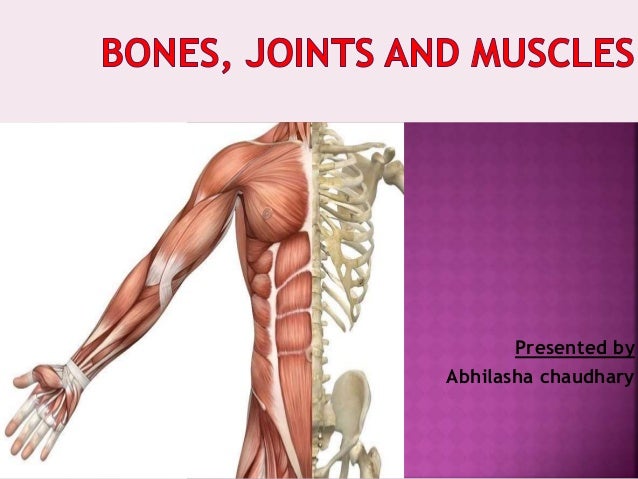
Bones Muscles And Joints from image.slidesharecdn.com Collection film x ray shoulder radiograph show shoulder dislocation and bone broken (neck of humerus fracture) from accident highlight on arrow point. Key facts about the main bones, joints and muscles of the body; Cartilage is found at the end of bones and is made of tough protein fibers. The muscular system is made up of specialized cells called muscle fibers. I highly recommend that muscle & bone ep and not just because i know the guys. Bones of the upper and lower limbs and the shoulder and pelvic girdles main joints: Neck bones and muscles pictures. Bones of the skull mandible occiput temporal bones parietal bones frontal bones maxilla bones.
Cartilage is found at the end of bones and is made of tough protein fibers.
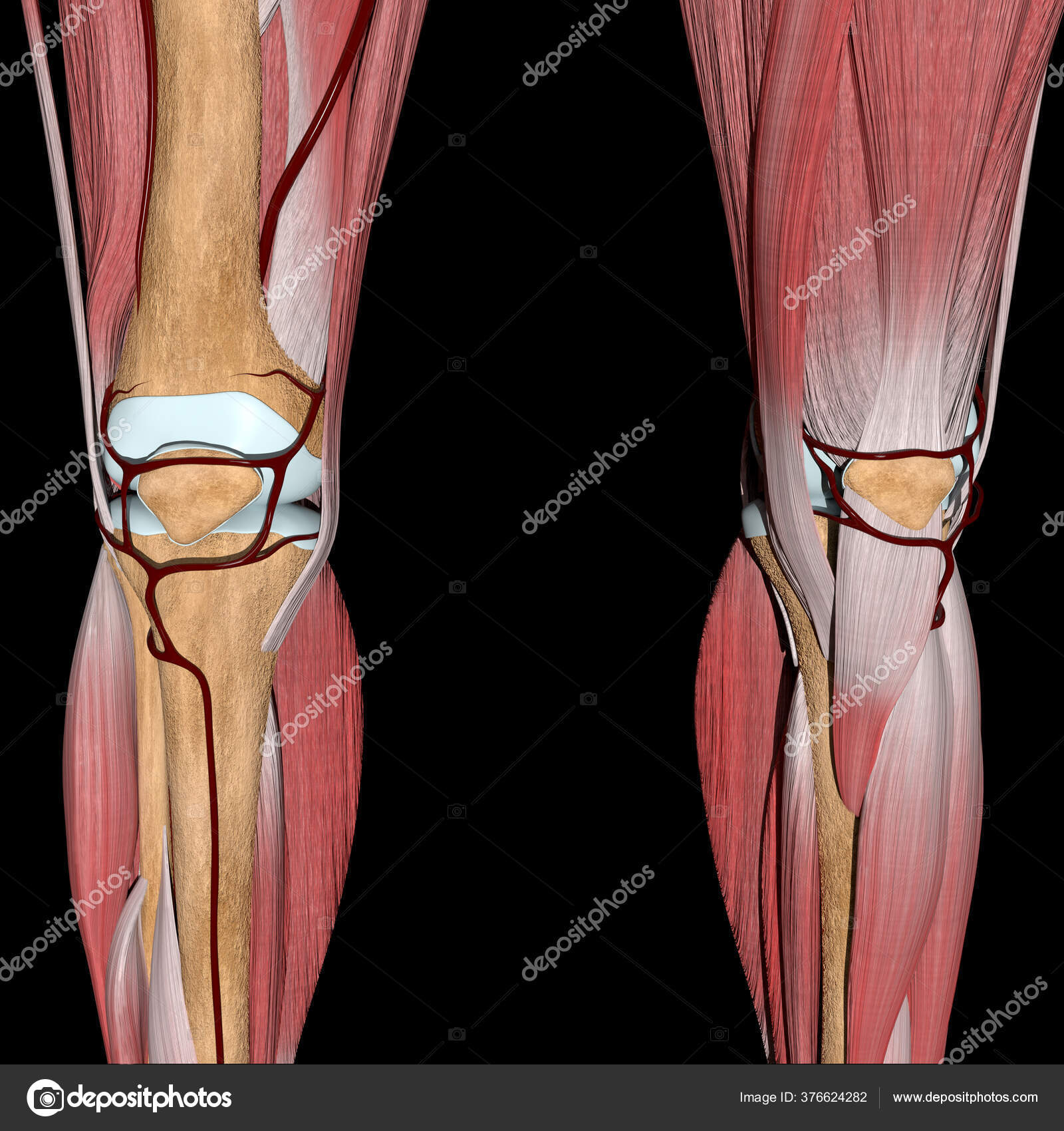
Secondarily, it protects the spinal cord (which is the extension of the brain) and all of the nerves that branch from the spinal cord. Download muscle bone stock photos. Picture of human head showing bones. 12 photos of the neck bones and muscles pictures. Collection film x ray shoulder radiograph show shoulder dislocation and bone broken (neck of humerus fracture) from accident highlight on arrow point. The calcaneus (heel bone) is the largest bone in the foot. I highly recommend that muscle & bone ep and not just because i know the guys. Human body anatomy muscles stock photos and images. Key facts about the main bones, joints and muscles of the body; The image below shows the bones of the hand from the back side. The knee joint is a complex structure that involves bones, tendons, ligaments, muscles, and other structures for normal function. Striated just like cardiac muscle, these skeletal muscle fibers are very strong. Related posts of neck bones and muscles pictures bone on you arm diagram.
Collection film x ray shoulder radiograph show shoulder dislocation and bone broken (neck of humerus fracture) from accident highlight on arrow point. Neck bones and muscles pictures. Secondarily, it protects the spinal cord (which is the extension of the brain) and all of the nerves that branch from the spinal cord. When there is damage to one of the structures that surround the knee joint, this can lead to discomfort and disability. Bones of the skull mandible occiput temporal bones parietal bones frontal bones maxilla bones.
Bones And Muscles Images Royalty Free Stock Bones And Muscles Photos Pictures Depositphotos from st3.depositphotos.com Collection film x ray shoulder radiograph show shoulder dislocation and bone broken (neck of humerus fracture) from accident highlight on arrow point. There are around 650 skeletal muscles within the typical human body. Neck bones and muscles pictures. The calcaneus (heel bone) is the largest bone in the foot. It looks like ivory and is extremely strong. *cries* a request asking how to draw serratus a. Pictures of muscles and bones. This anatomical atlas was especially designed for a specific public (radiologists.
Muscle tissue is made up of bands of cells that contract and allow bodies to move.
As well as some basic images of disc pathology and stylised facet joint motion. Skeletal muscle cells form when many smaller progenitor cells lump themselves together to form long, straight, multinucleated fibers. This anatomical atlas was especially designed for a specific public (radiologists. Collection film x ray shoulder radiograph show shoulder dislocation and bone broken (neck of humerus fracture) from accident highlight on arrow point. The basics on muscles, bones, and joints. Pictures of muscles and bones. Lever infographic diagram showing parts and types including fulcrum load and effort with examples of human body joints bones and muscles daily lives for physics science education. Neck bones and muscles pictures. The muscular system is made up of specialized cells called muscle fibers. It looks like ivory and is extremely strong. Begins with the structural characteristics of bones and muscle mass. The flexor contracts to bend a limb at a joint. Most skeletal muscles are attached to two bones across a joint, so the muscle serves to move parts of those bones closer to each other.
This is a table of skeletal muscles of the human anatomy. The tarsal bones are found near the. Browse 4,015 shoulder bone stock photos and images available, or search for pork shoulder bone to find more great stock photos and pictures. The purpose of the spine is to support the body so that we can stand upright. Picture of human head showing bones.
Bones And Muscles Theschoolrun from www.theschoolrun.com The basics on muscles, bones, and joints. Find out how the musculoskeletal system functions — and which medical. Your bones, joints, and muscles make up your muscular and skeletal systems. Neck bones and muscles pictures 12 photos of the neck bones and muscles pictures , bone. When there is damage to one of the structures that surround the knee joint, this can lead to discomfort and disability. Almost every muscle constitutes one part of a pair of identical bilateral muscles, found on both sides, resulting in approximately 320 pairs of muscles, as presented in this article. September 23, 2019 edited by dr. As well as some basic images of disc pathology and stylised facet joint motion.
Neck bones and muscles pictures.
Images provided by the nemours. See more ideas about anatomy, thoracic, basic image. *cries* a request asking how to draw serratus a. 12 photos of the neck bones and muscles pictures. Lever infographic diagram showing parts and types including fulcrum load and effort with examples of human body joints bones and muscles daily lives for physics science education. This anatomical atlas was especially designed for a specific public (radiologists. There are around 650 skeletal muscles within the typical human body. Secondarily, it protects the spinal cord (which is the extension of the brain) and all of the nerves that branch from the spinal cord. The flexor contracts to bend a limb at a joint. Picture of human head showing bones. Learning anatomy requires more than pictures and labels it requires a. Begins with the structural characteristics of bones and muscle mass. Muscles, tendons, and ligaments run along the surfaces of the feet, allowing the complex movements needed for motion and balance.


Komentar
Posting Komentar